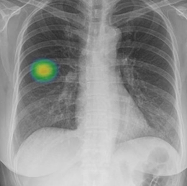
As per a study, the algorithm performs with higher efficiency and effectiveness compared to the existing methods.
A team of doctors, scientists, and researchers have collaborated to develop an AI model that can precisely detect cancer, potentially accelerating the diagnosis and treatment of the disease.
With nearly 10 million deaths each year, cancer is a major cause of mortality across the globe, accounting for nearly one in six deaths, as reported by the World Health Organization. Nonetheless, timely detection and prompt treatment can cure the disease in many cases.
The algorithm, designed by professionals from Imperial College London, the Institute of Cancer Research, London, and the Royal Marsden NHS Foundation Trust, can accurately distinguish cancerous growths from non-cancerous ones spotted on CT scans.
A study published in the Lancet’s eBioMedicine journal has revealed that an algorithm performs with higher efficiency and effectiveness than current methods for detecting cancer. The algorithm was developed by experts from the Royal Marsden NHS foundation trust, Imperial College London, and the Institute of Cancer Research, London.

Dr Benjamin Hunter, a clinical oncology registrar at the Royal Marsden and a clinical research fellow at Imperial, stated that the algorithm could improve early detection and potentially make cancer treatment more successful by highlighting high-risk patients and fast-tracking them to earlier intervention.
The team used radiomics to extract essential information from CT scans of around 500 patients with large lung nodules and develop an AI algorithm. The model was then tested to identify if it could accurately distinguish cancerous nodules. The study used the area under the curve (AUC) measure to determine the effectiveness of the model at predicting cancer. The results showed that the AI model could identify the risk of cancer in each nodule with an AUC of 0.87, outperforming the Brock score, a current clinic test, which scored 0.67.

The next step is to test the technology on patients with large lung nodules in clinic to see if it can accurately predict their risk of lung cancer. The AI model may also assist physicians to reach more timely choices regarding patients with abnormal growths classified as medium-risk.
The researchers, supported by the Royal Marsden Cancer Charity, the National Institute for Health and Care Research, RM Partners, and Cancer Research UK, stress that more testing must be done before the model can be implemented in healthcare systems. The Libra study is still in its early stages.
Richard Lee, the study’s chief investigator, explained that lung cancer is the leading cause of cancer mortality globally, causing one-fifth of cancer deaths in the United Kingdom. Early detection allows for more successful treatment, yet current data suggest that more than 60% of lung cancer cases in England are detected at stage three or four. Lee stated that people diagnosed with lung cancer at the earliest stage are much more likely to survive for five years compared with those whose cancer is caught late. The development of the radiomics model specifically focused on large lung nodules could one day support clinicians in identifying high-risk patients.
SOURCE: THE GUARDIAN AND NEWS AGENCIES


